


Knowledge Base
Immune Modulation & Inflammatory Response

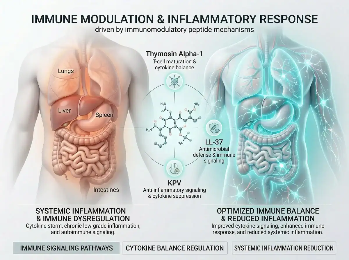
Immune Modulation & Inflammatory Response: The Science Behind Peptides That Influence Immune Signaling and Cytokine Balance
How immunomodulatory peptides are being studied for their potential roles in immune enhancement, antimicrobial defense, and inflammatory regulation
The Immune System: Guardian and Potential Adversary
The immune system of the human body represents one of biology's most sophisticated achievements—a distributed network of cells, tissues, and signaling molecules capable of distinguishing self from non-self, eliminating pathogens, and maintaining tissue homeostasis.
Yet this same system can become dysregulated, contributing to disease rather than preventing it.
The dual nature of immunity:
- Too weak: Increased susceptibility to infections, cancer, chronic disease
- Too strong: Autoimmune conditions, chronic inflammation, tissue damage
- Misdirected: Allergies, hypersensitivities, inflammatory disorders
Chronic inflammation—now recognized as a driver of aging itself ("inflammaging")—connects immune dysfunction to virtually every major age-related disease, from cardiovascular disease to neurodegeneration to metabolic syndrome.
The central question for immunological research: Can immune function be therapeutically optimized—enhanced where deficient, modulated where excessive, and balanced where dysregulated?
Emerging research into immunomodulatory peptides suggests promising pathways. Four compounds in particular—Thymosin Alpha-1, Thymulin, LL-37, and KPV—represent distinct approaches to immune modulation, each with unique mechanisms for influencing immune cell function, cytokine production, and inflammatory responses.
Understanding Immune Modulation: Beyond Simple Enhancement
Effective immune modulation requires understanding that the goal is rarely simple "boosting." Instead, optimal immune function involves balanced, context-appropriate responses.
Balanced Immune Activation
| State | Characteristics | Consequences |
|---|---|---|
| Hypoactive | Reduced T-cell function, poor pathogen recognition | Infections, cancer progression |
| Hyperactive | Excessive inflammation, cytokine overproduction | Tissue damage, autoimmunity |
| Balanced | Appropriate responses, proper resolution | Health, homeostasis |
Key Immune Regulatory Systems
Innate Immunity: First-line defense including macrophages, neutrophils, and NK cells. Pattern recognition receptors (Toll-like receptors) enable rapid, non-specific response and initiate adaptive immunity.
Adaptive Immunity: Antigen-specific responses through T cells and B cells. Provides immunological memory with slower but precisely targeted responses for long-term protection.
Regulatory Mechanisms: Regulatory T cells (Tregs) suppress excessive responses. Anti-inflammatory cytokines (IL-10, TGF-β) balance pro-inflammatory signals. Resolution pathways actively terminate inflammation.
The peptides examined in this article influence these systems through different mechanisms—offering researchers tools for understanding and potentially modifying immune responses.
The Cytokine Network: Immune Communication
What Are Cytokines?
Cytokines are small signaling proteins that mediate communication between immune cells and coordinate inflammatory responses. Understanding key cytokines is essential for appreciating immunomodulatory peptide mechanisms.
Pro-inflammatory Cytokines:
| Cytokine | Primary Sources | Key Functions |
|---|---|---|
| TNF-α | Macrophages, T cells | Activates inflammation, induces fever, promotes apoptosis |
| IL-1β | Macrophages, monocytes | Fever, inflammation, T-cell activation |
| IL-6 | Many cell types | Acute phase response, B-cell differentiation |
| IFN-γ | T cells, NK cells | Macrophage activation, antiviral responses |
| IL-8 (CXCL8) | Macrophages, epithelial cells | Neutrophil chemotaxis |
Anti-inflammatory Cytokines:
| Cytokine | Primary Sources | Key Functions |
|---|---|---|
| IL-10 | Tregs, macrophages | Suppresses pro-inflammatory cytokines |
| TGF-β | Many cell types | Immune suppression, tissue repair |
| IL-4 | Th2 cells | Alternative macrophage activation |
The Cytokine Storm Problem
When pro-inflammatory cytokine production becomes uncontrolled, the resulting "cytokine storm" can cause severe tissue damage—as observed in severe COVID-19, sepsis, and certain autoimmune conditions. Effective immunomodulation aims to support appropriate cytokine responses while preventing dangerous overproduction.
Thymosin Alpha-1: The Thymic Immunomodulator
Origins and Structure
Thymosin Alpha-1 (Tα1), also known as thymalfasin, is a 28-amino-acid peptide naturally produced by the thymus gland—the organ responsible for T-cell maturation and education.
The peptide has the sequence Ac-SDAAVDTSSEITTKDLKEKKEVVEEAEN. It is N-terminally acetylated and endogenously produced throughout life, declining with age. The synthetic version is identical to the natural peptide.
The thymus undergoes progressive involution (shrinkage) with age, leading to reduced Tα1 production and contributing to age-related immune decline (immunosenescence).
Thymosin Alpha-1
59,90 € Vialincl. VAT
plus Shipping Costs
Delivery time: 1–6 Working Days
Mechanism of Action
Thymosin Alpha-1 operates through multiple immunomodulatory pathways, earning it the description of having "pleiotropic" (multiple) effects.
T-Cell Enhancement: Tα1 promotes T-cell development, differentiation, and function. It stimulates differentiation of precursor stem cells into mature T cells, increases CD4+ helper T cells, CD8+ cytotoxic T cells, and CD3+ populations, balances CD4/CD8 ratios, enhances T-cell receptor expression, and promotes IL-2 receptor upregulation enabling T-cell activation.
Dendritic Cell Activation: Dendritic cells (DCs) are professional antigen-presenting cells that bridge innate and adaptive immunity. Tα1 acts via Toll-like receptors (TLR2, TLR9) on myeloid and plasmacytoid DCs, activates NF-κB and MAPK signaling pathways, enhances antigen presentation, and promotes cytokine production for T-cell priming.
Natural Killer (NK) Cell Stimulation: NK cells provide rapid cytotoxic responses against infected or malignant cells. Tα1 increases NK cell activity and cytotoxicity, enhances ability to directly kill virally infected cells, and supports tumor surveillance.
Cytokine Modulation: Tα1 influences cytokine networks bidirectionally. It enhances IFN-γ, IL-2, and IL-3 to support immune activation while promoting IL-10-producing regulatory T cells to provide feedback inhibition. This prevents excessive cytokine release, protecting against cytokine storms.
This dual action—enhancing immune responses while preventing overactivation—represents Tα1's key advantage as an immunomodulator rather than simple immune stimulant.
Oxidative Stress Protection: Tα1 demonstrates antioxidant properties, reducing reactive oxygen species (ROS) in immune cells, protecting against oxidative damage during immune activation, and supporting cellular function under inflammatory conditions.
Clinical Applications and Research
Tα1 has been studied extensively in various clinical contexts.
Infectious Diseases: Chronic hepatitis B and C (primary approved indication), HIV/AIDS (adjunctive therapy), severe bacterial infections, and viral infections including COVID-19 (investigational).
Oncology: Adjunctive cancer immunotherapy, reducing chemotherapy-induced immunosuppression, enhancing vaccine responses in cancer patients, and supporting immune surveillance.
Immunodeficiency: Age-related immune decline, post-transplant immune recovery, and chemotherapy-induced immune suppression.
Recent Research Developments (2024-2025)
Cancer Immunotherapy Combinations: A 2024 study demonstrated that Tα1 combined with anti-PD-1 immunotherapy significantly improves outcomes in hepatocellular carcinoma (HCC) patients post-hepatectomy. The combination group achieved 1- and 2-year recurrence-free survival rates of 98.4% and 80.2%, compared to 86.2% and 65.8% for anti-PD-1 alone, and 49.2% and 24.6% for control groups (p < 0.001). Multivariable analysis showed the combination treatment significantly reduced recurrence and mortality risk.
For melanoma, Tα1 combined with chemotherapy (dacarbazine) increased the response rate threefold in stage IV patients compared to chemotherapy alone.
Vaccine Response Enhancement: A Phase 1 study is currently testing different treatment regimens of Tα1 before SARS-CoV-2 mRNA vaccination in moderately to severely immunocompromised patients, including those with active cancer treatment or hematologic malignancies. The study measures safety, tolerability, and antibody responses over 24-52 weeks.
COVID-19 Research: Clinical trials evaluated Tα1 for hospitalized COVID-19 patients with lymphocytopenia. A retrospective analysis from China showed that patients treated with Tα1 demonstrated improved lymphocyte subsets and significantly reduced mortality (from 30% to 11%, p = 0.04). Treatment involves daily subcutaneous injections of 1.6 mg for one week.
Sepsis Outcomes—Important Update: A large 2025 placebo-controlled trial (TESTS) with over 1,000 participants found that Tα1 showed no clear evidence of reducing 28-day all-cause mortality in adults with sepsis. The trial enrolled 1,089 participants and found 23.4% mortality in the Tα1 group versus 24.1% in the placebo group (hazard ratio 0.99, 95% CI 0.77–1.27, p = 0.93). This contradicts earlier smaller trials suggesting Tα1's efficacy in sepsis, highlighting the importance of large, well-designed trials.
Regulatory Status
Approved in 35+ Countries: China, Italy, Russia, and others. Primary indication is chronic hepatitis B and C. Additional approved uses vary by country.
United States: Not FDA approved for any indication. Received Orphan Drug designation for certain conditions. Available through research and compounding. FDA has included Tα1 in 2024 guidance on peptides of concern for compounding.
European Union: Not centrally approved by EMA. Individual country approvals vary. Research compound in most EU nations.
Safety Profile
Tα1 demonstrates an excellent safety record spanning over four decades.
Reported Side Effects: Mild, temporary injection site reactions (most common). Rare: mild flu-like symptoms. No serious adverse effects in clinical trials.
Contraindications/Cautions: Pregnancy/breastfeeding (insufficient data). Severe autoimmune conditions (may modulate immune responses unpredictably). Organ transplant recipients (theoretical concern for rejection).
Long-term Safety: Clinical use since 1979 with well-documented safety profile. Multiple trials in hepatitis, HIV, and cancer patients show favorable tolerability.
Thymulin: The Zinc-Dependent Thymic Hormone
Origins and Structure
Thymulin (formerly known as Facteur Thymique Sérique or FTS) is a nonapeptide (9 amino acids) produced by thymic epithelial cells. It represents one of the major thymic hormones alongside Thymosin Alpha-1.
Key characteristics:
- Sequence: Pyr-Ala-Lys-Ser-Gln-Gly-Gly-Ser-Asn (pyroglutamic acid at N-terminus)
- Absolutely requires zinc for biological activity
- Forms a 1:1 complex with Zn²⁺
- The zinc-bound form is the only active form
- Production declines with age (parallel to thymic involution)
The Zinc Requirement: Thymulin is unique among thymic peptides in its absolute dependence on zinc. Without zinc bound, thymulin has no biological activity. This creates a direct link between zinc status and thymulin function—and by extension, between zinc status and thymic-mediated immunity.
Thymalin / Thymulin
39,90 €incl. VAT
plus Shipping Costs
Delivery time: 1–6 Working Days
Mechanism of Action
Thymulin operates primarily through T-cell modulation and neuroendocrine interactions.
T-Cell Differentiation and Maturation: Thymulin promotes the differentiation of immature T-cell precursors into mature, functional T cells. It enhances expression of T-cell markers (CD3, CD4, CD8), supports thymic education (self vs. non-self recognition), and maintains T-cell homeostasis.
Cytokine Modulation: Thymulin influences cytokine production by T cells and other immune cells. It modulates IL-2, IL-6, and other cytokines, supports balanced Th1/Th2 responses, and may help regulate inflammatory responses.
Neuroendocrine Integration: Thymulin participates in bidirectional communication between the immune and neuroendocrine systems. It interacts with the hypothalamic-pituitary-adrenal axis, influences glucocorticoid sensitivity, and may affect stress responses and immune function integration.
Anti-inflammatory Properties: Research suggests thymulin has anti-inflammatory effects, reducing inflammatory cytokine production in certain models, potentially protecting against excessive inflammation, and supporting tissue homeostasis.
The Thymulin-Zinc-Aging Connection
The relationship between thymulin, zinc, and aging creates an important convergence:
Age-Related Decline:
- Thymulin production decreases with thymic involution
- Circulating thymulin levels drop significantly with age
- By age 60, thymulin may be nearly undetectable in serum
Zinc Status in Aging:
- Zinc deficiency is common in older populations
- Even marginal zinc deficiency impairs thymulin activity
- Zinc supplementation can restore thymulin function in zinc-deficient individuals
Immunosenescence Connection:
- Declining thymulin contributes to T-cell dysfunction
- Reduced naive T-cell production
- Impaired immune responses to new antigens
- Reduced vaccine efficacy in elderly populations
Research Applications
Immunosenescence: Thymulin research has focused on reversing age-related immune decline. Zinc supplementation studies show restoration of thymulin activity in deficient elderly subjects. Animal models demonstrate improved T-cell function with thymulin administration.
Autoimmune and Inflammatory Conditions: Limited research suggests potential applications in autoimmune conditions, with thymulin showing anti-inflammatory effects in some models.
Neuroimmune Interactions: Research explores thymulin's role in brain-immune communication, stress responses, and neuroinflammation.
Current Development Status
Regulatory Status:
- Not FDA approved
- Not EMA approved
- Research compound only
- No registered therapeutic clinical trials
Research Stage:
- Primarily preclinical and mechanistic studies
- Some human studies on zinc-thymulin restoration
- No advanced clinical development for therapeutic use
Safety Profile
Available Evidence: Very limited human safety data exists for exogenous thymulin administration. Endogenous thymulin is a naturally occurring hormone.
Considerations:
- Zinc status affects activity (zinc supplementation may be required)
- Theoretical concerns about immune modulation in autoimmune conditions
- Limited data overall
LL-37: The Antimicrobial Defender
Origins and Structure
LL-37 is the only human cathelicidin antimicrobial peptide—a 37-amino-acid peptide (hence "LL-37," beginning with two leucines) derived from the precursor protein hCAP18.
The sequence is LLGDFFRKSKEKIGKEFKRIVQRIKDFLRNLVPRTES. The peptide has an α-helical structure in membrane environments and is produced by neutrophils, epithelial cells, macrophages, and other immune cells. Expression is induced by vitamin D, infection, and inflammation.
LL-37 represents an ancient component of innate immunity, with antimicrobial peptides (AMPs) found throughout the animal kingdom as first-line defenders against pathogens.
Mechanism of Action
LL-37 operates through multiple mechanisms spanning antimicrobial activity, immune modulation, and tissue repair.
Direct Antimicrobial Activity: LL-37 kills pathogens through membrane disruption. Bacterial killing occurs through forming pores in bacterial membranes, causing cytoplasmic leakage. The peptide demonstrates selectivity, targeting anionic microbial membranes (LPS, teichoic acids) while sparing mammalian cells. Broad spectrum activity includes Gram-positive, Gram-negative bacteria, fungi, and some viruses. Biofilm disruption occurs at low concentrations (0.5 μg/ml).
| Pathogen Type | Examples | Mechanism |
|---|---|---|
| Gram-negative | P. aeruginosa, E. coli | LPS binding, membrane pore formation |
| Gram-positive | S. aureus, S. pneumoniae | Teichoic acid binding, membrane disruption |
| Fungi | Candida albicans, Aspergillus | Membrane permeabilization |
| Viruses | Influenza, HSV, HIV (partial) | Envelope disruption, entry inhibition |
Endotoxin Neutralization: One of LL-37's most important functions is neutralizing bacterial endotoxins. The peptide binds directly to lipopolysaccharide (LPS) and lipoteichoic acid (LTA), prevents endotoxin-induced inflammatory cascades, reduces TNF-α and other pro-inflammatory cytokine release, and may protect against sepsis-related inflammation.
Immune Cell Modulation: LL-37 influences multiple immune cell types. For chemotaxis, it attracts neutrophils, monocytes, and T cells to infection sites, induces MCP-1/CCL2 and CXCL8 chemokine production, and coordinates immune cell recruitment. For macrophage activation, it promotes antimicrobial activity, influences macrophage polarization, and enhances phagocytosis. For dendritic cell effects, it modulates DC maturation, influences antigen presentation, and affects T-cell priming.
Receptor-Mediated Signaling: LL-37 activates multiple cellular receptors: FPRL-1 (Formyl Peptide Receptor-Like 1) for chemotaxis and immune activation, P2X7 for ATP-dependent inflammation and NLRP3 inflammasome, EGFR for wound healing, and TLRs for Toll-like receptor modulation.
Wound Healing and Tissue Repair: Beyond antimicrobial functions, LL-37 promotes tissue regeneration. It stimulates keratinocyte and fibroblast proliferation, promotes cell migration for re-epithelialization, induces angiogenesis (new blood vessel formation), and supports barrier function restoration.
Research Applications
LL-37 is being studied for various applications.
Infectious Disease: Antimicrobial therapy (particularly antibiotic-resistant infections), anti-biofilm strategies, wound infection prevention, and sepsis management.
Wound Healing: Chronic wound treatment, burn healing, and surgical wound management.
Inflammatory Conditions: Modulating excessive inflammation, endotoxin-related pathology, and mucosal immunity.
Cancer Research: Complex, context-dependent effects exist. LL-37 may be pro-tumorigenic in some cancers (EGFR activation) and anti-tumorigenic in others (direct cytotoxicity). Careful evaluation by cancer type is required.
Current Development Status
Regulatory Status: Not FDA approved for any therapeutic indication. No EMA approval. Remains investigational/research compound. FDA has noted concerns about compounded LL-37 products.
Research Stage: Primarily preclinical studies. Limited human clinical data. Topical formulations under investigation. Systemic use faces delivery challenges.
Safety Profile
Available Safety Data: Generally well-tolerated in available studies. Excellent safety profile in animal and limited human research.
Reported Effects: Injection site reactions (redness, irritation), transient flu-like symptoms, temporary fatigue, mild GI upset.
Concerns: At high (μM) concentrations, can damage host cell membranes. Theoretical concerns about tumor promotion in certain contexts. FDA has noted concerns regarding male fertility in nonclinical studies. Long-term human safety data is limited.
KPV: The Anti-Inflammatory Tripeptide
Origins and Structure
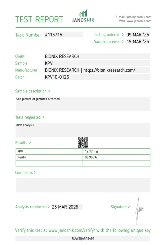
KPV is a tripeptide with the sequence Lys-Pro-Val (Lysine-Proline-Valine)—representing the C-terminal fragment of alpha-melanocyte-stimulating hormone (α-MSH).
The peptide consists of only 3 amino acids (the smallest peptide in this review). It is derived from α-MSH positions 11-13, retains anti-inflammatory activity without melanocortin receptor binding, and has a stable, simple structure enabling various delivery routes.
α-MSH is a 13-amino-acid peptide with diverse functions including pigmentation, appetite regulation, and immune modulation. KPV retains the anti-inflammatory properties while eliminating effects on melanin production and appetite.
KPV
34,90 € Vialincl. VAT
plus Shipping Costs
Delivery time: 1–6 Working Days
Mechanism of Action
KPV operates through mechanisms distinct from Thymosin Alpha-1, Thymulin, and LL-37.
PepT1-Mediated Uptake: KPV's primary mechanism involves the peptide transporter PepT1. The peptide is actively transported into intestinal epithelial cells via hPepT1, achieves high intracellular concentrations, and this uptake is essential for anti-inflammatory effects. Blocking PepT1 abolishes KPV's activity.
NF-κB Inhibition: Once inside cells, KPV inhibits the master inflammatory transcription factor NF-κB. It blocks NF-κB activation and nuclear translocation, reduces transcription of pro-inflammatory genes, decreases production of IL-8, TNF-α, and other inflammatory mediators, and operates independently of melanocortin receptor activation.
MAPK Pathway Modulation: KPV also affects mitogen-activated protein kinase signaling. It inhibits MAPK pathway activation, reduces inflammatory gene expression, and complements NF-κB inhibition.
IL-1β Antagonism: KPV demonstrates specific effects on IL-1β signaling. It antagonizes IL-1β activity, reduces IL-1β-induced inflammatory responses, and may protect against inflammasome-driven inflammation.
Macrophage Polarization: KPV influences macrophage phenotype. It promotes M2 (anti-inflammatory) macrophage polarization, reduces M1 (pro-inflammatory) phenotype, and shifts immune response toward resolution.
Barrier Function Support: In intestinal applications, KPV supports epithelial integrity. It upregulates tight junction proteins (occludin, ZO-1), restores barrier function, reduces intestinal permeability ("leaky gut"), and enhances mucosal healing.
Research Applications
KPV research has focused primarily on inflammatory conditions.
Inflammatory Bowel Disease (IBD): DSS-induced colitis models show significant improvement. TNBS colitis responds to KPV treatment. The peptide reduces disease activity, cytokine levels, and histological damage. Oral administration is effective in animal models.
Gut Health: Intestinal barrier repair, mucosal healing, and microbiome support (indirect effects).
Skin Inflammation: Potential applications in inflammatory skin conditions, wound healing support, and barrier function restoration.
Systemic Inflammation: General anti-inflammatory applications that may complement other anti-inflammatory strategies.
Recent Research Developments (2024-2025)
Advanced Delivery Systems: Lipid nanocapsule systems (LNC-Ted HAKPV) deliver KPV with hyaluronic acid (HA), promoting GLP-2 production for intestinal repair, modulating CD44/TLR4 pathways for anti-inflammation, and enabling redox-responsive release in inflamed tissue.
HA-KPV conjugation enhances colon-specific targeting, reducing systemic exposure and supporting microbiota homeostasis.
ROS-Responsive Prodrugs: New formulations release KPV specifically in inflamed tissue where reactive oxygen species are elevated. These enhanced formulations show superior efficacy over free KPV.
Current Development Status
Regulatory Status: Not FDA approved for any indication. Not EMA approved. Research compound only. No registered clinical trials for therapeutic use.
Research Stage: Primarily preclinical (in vitro, animal models). Strong mechanistic understanding. Limited human data. Novel delivery systems under development.
Safety Profile
Available Safety Data: KPV demonstrates an excellent safety profile in available research.
Reported Effects: Very rare adverse reactions. Well-tolerated in animal models. No specific side effects documented in available literature.
Advantages: Tripeptide structure (simple, low immunogenicity risk). Targeted anti-inflammatory action. Does not cause broad immunosuppression. Oral bioavailability (unlike many peptides).
Limitations: No human clinical trial safety data. Long-term effects unknown. Potential for individual variability.
The NAD+-Sirtuin-Immune Connection
How NAD+ Influences Immune Function
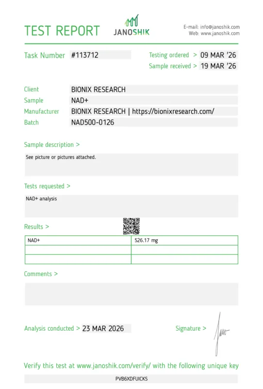
While Thymosin Alpha-1, Thymulin, LL-37, and KPV directly target immune cells and inflammatory pathways, NAD+ (nicotinamide adenine dinucleotide) influences immunity through a parallel metabolic pathway—the sirtuins.
The NAD+-Sirtuin-Inflammation Axis:
NAD+ is essential for sirtuin function. Sirtuins (especially SIRT1, SIRT6) are NAD+-dependent enzymes. Without adequate NAD+, sirtuin activity diminishes.
SIRT1 inhibits NF-κB by deacetylating the p65 subunit of NF-κB, reducing transcription of pro-inflammatory genes, and decreasing TNF-α, IL-1β, and IL-6 production.
SIRT6 modulates inflammation by regulating inflammatory gene expression, influencing macrophage function, and supporting immune cell metabolism.
NAD+ decline promotes inflammation. Age-related NAD+ decline correlates with chronic inflammation. Senescent cells increase CD38 expression, consuming NAD+. This creates a feedback loop: inflammation leads to NAD+ depletion, which causes more inflammation.
NAD+
59,90 € Vialincl. VAT
plus Shipping Costs
Delivery time: 1–6 Working Days
Research Evidence:
| Finding | Implication |
|---|---|
| NAD+ precursor (NR) reduces pro-inflammatory cytokines | Supporting NAD+ may dampen chronic inflammation |
| Chronic inflammation depletes NAD+ via CD38 | Inflammation and NAD+ decline are interconnected |
| SIRT1 activation reduces inflammatory markers | NAD+-dependent sirtuins are key regulators |
| Inflammaging correlates with NAD+ decline | Maintaining NAD+ may support immune homeostasis |
Important Nuance: The relationship between NAD+ and inflammation is context-dependent. In chronic inflammation, NAD+ support generally reduces inflammatory markers. In acute infection, some NAD+ pathways support necessary immune activation. The goal is balanced immune function, not simple suppression.
Integrating NAD+ with Immunomodulatory Peptides
A systems approach to immune modulation might consider multiple pathways:
| Target | Compound | Primary Effect |
|---|---|---|
| T-cell/DC enhancement | Thymosin Alpha-1 | Adaptive immunity support |
| T-cell maturation | Thymulin (+ zinc) | Thymic function support |
| Antimicrobial defense | LL-37 | Innate immunity, pathogen killing |
| NF-κB inhibition | KPV | Direct anti-inflammatory |
| Sirtuin activation | NAD+ precursors | Metabolic-immune regulation |
These pathways are complementary rather than redundant, potentially offering different approaches for different immune challenges.
Comparative Analysis: Four Approaches to Immune Modulation
| Characteristic | Thymosin Alpha-1 | Thymulin | LL-37 | KPV |
|---|---|---|---|---|
| Structure | 28 amino acids | 9 amino acids | 37 amino acids | 3 amino acids |
| Origin | Thymus gland | Thymus epithelial cells | Neutrophils, epithelial cells | α-MSH fragment |
| Cofactor Required | None | Zinc (essential) | None | None |
| Primary Target | T cells, DCs, NK cells | T-cell precursors | Pathogens, immune cells | NF-κB pathway |
| Main Function | Immune enhancement/balance | T-cell maturation | Antimicrobial, immunomodulation | Anti-inflammatory |
| Receptor Mechanism | TLR2, TLR9 | Specific thymulin receptors | FPRL-1, P2X7, EGFR | PepT1 transport |
| Cytokine Effects | ↑ IFN-γ, IL-2; promotes IL-10 | Modulates IL-2, IL-6 | Modulates TNF-α, chemokines | ↓ IL-8, TNF-α, IL-1β |
| Approved Uses | Hepatitis B/C (35+ countries) | None | None | None |
| Administration | Subcutaneous injection | Injection | Injectable, topical | Oral, injectable, topical |
| Safety Data | Extensive (40+ years) | Very limited | Limited | Very limited |
Complementary Applications
The four peptides address different aspects of immune dysfunction.
Thymosin Alpha-1 is best suited for immunodeficiency states, chronic infections, cancer adjunctive therapy, and age-related immune decline.
Thymulin is best suited for zinc deficiency-related immune dysfunction, supporting thymic function in aging, and T-cell maturation deficits.
LL-37 is best suited for antimicrobial applications, wound healing, biofilm-associated infections, and mucosal defense.
KPV is best suited for inflammatory bowel conditions, gut barrier dysfunction, localized inflammation, and anti-inflammatory support.
Safety Considerations and Current Limitations
Comparative Safety Profiles
| Peptide | Safety Evidence | Common Effects | Serious Concerns |
|---|---|---|---|
| Thymosin Alpha-1 | Extensive (decades of clinical use) | Injection site reactions | Autoimmune modulation (caution) |
| Thymulin | Very limited | Unknown | Zinc-dependent activity |
| LL-37 | Limited (mostly preclinical) | Injection reactions, flu-like symptoms | Tumor concerns, fertility (nonclinical) |
| KPV | Very limited (animal studies) | None documented | Unknown long-term effects |
FDA Regulatory Actions
The FDA's 2024 guidance on pharmacy compounding addressed several peptides. Thymosin Alpha-1 is included in the list of peptides with safety concerns for compounding. LL-37 has noted concerns regarding nonclinical safety findings. KPV and Thymulin are not specifically addressed but remain unapproved.
These regulatory actions reflect concern about quality, purity, and safety of compounded peptide products rather than necessarily indicating inherent dangers of the compounds themselves when properly manufactured.
Quality and Purity Concerns
As with all research peptides, source quality significantly impacts safety. Pharmaceutical-grade products (where available) offer highest quality assurance. Compounded products vary in quality control. Research-grade peptides may have lower purity standards. Contamination and degradation risks exist with improper handling.
The Regulatory Landscape
Current Status Summary
| Peptide | FDA Status | EMA Status | Other Approvals |
|---|---|---|---|
| Thymosin Alpha-1 | Not approved; orphan drug designation | Not centrally approved | Approved in 35+ countries |
| Thymulin | Not approved | Not approved | None |
| LL-37 | Not approved | Not approved | None |
| KPV | Not approved | Not approved | None |
Research vs. Therapeutic Use
All four peptides represent different stages of development. Thymosin Alpha-1 is most clinically advanced, approved in many countries with a strong safety record. Thymulin is primarily a research tool with limited therapeutic development. LL-37 has promising preclinical data, limited human studies, and delivery challenges. KPV has strong mechanistic understanding, primarily animal data, and novel delivery systems emerging.
Summary: The Future of Immune Modulation
Immunomodulatory peptides represent a sophisticated approach to supporting immune function—moving beyond simple "immune boosting" toward balanced, context-appropriate immune responses.
Key Insights:
Immune modulation requires balance—enhancing function where deficient while preventing overactivation that causes tissue damage.
Four distinct mechanisms offer complementary approaches. Thymosin Alpha-1 enhances adaptive immunity through T-cell and DC activation. Thymulin supports T-cell maturation in a zinc-dependent manner. LL-37 provides innate antimicrobial defense and wound healing support. KPV offers targeted anti-inflammatory action through NF-κB inhibition.
The NAD+-sirtuin axis connects cellular energy metabolism to immune regulation, providing another pathway for supporting immune homeostasis.
Clinical development varies significantly. Thymosin Alpha-1 has decades of clinical use and approval in multiple countries, with recent research supporting combination immunotherapy approaches. Thymulin, LL-37, and KPV remain primarily research compounds.
Safety profiles differ. Thymosin Alpha-1 has extensive safety documentation. Thymulin, LL-37, and KPV have limited human safety data.
For researchers and those following developments in immunology and longevity science, immunomodulatory peptides offer valuable tools for understanding the complex interplay between immune function, inflammation, and aging.
The immune system's dual role—as both guardian and potential source of pathology—makes precise modulation one of medicine's most challenging and important frontiers.
Frequently Asked Questions: Immunomodulatory Peptides & Inflammatory Response
General Questions About Immune Modulation
Q: What is immune modulation and how is it different from immune boosting?
Immune modulation refers to therapeutic adjustment of immune function toward optimal balance—which may involve enhancement, suppression, or redirection depending on the underlying condition.
Key distinction:
- Immune boosting: Simple enhancement of immune activity (may worsen autoimmune conditions)
- Immune modulation: Intelligent adjustment toward appropriate responses (enhances where weak, calms where overactive)
The immune system can cause disease through both underactivity (infections, cancer) and overactivity (autoimmunity, chronic inflammation). Effective immunomodulation addresses the specific dysfunction rather than simply "boosting" all immune responses.
Thymosin Alpha-1 exemplifies true immunomodulation—it enhances T-cell function while also promoting regulatory T cells that prevent excessive inflammation.
Q: What is inflammaging and why does it matter?
Inflammaging describes the chronic, low-grade inflammation that develops with aging and contributes to age-related diseases. It represents a key connection between immune dysfunction and the aging process.
Characteristics: Elevated baseline inflammatory markers (IL-6, TNF-α, CRP), present without acute infection or injury, persists over time, accelerates tissue aging and disease.
Contributing factors: Senescent cell accumulation (SASP secretion), gut barrier dysfunction ("leaky gut"), chronic infections (CMV, etc.), obesity and metabolic dysfunction, declining immune regulation.
Disease connections: Inflammaging contributes to cardiovascular disease, neurodegeneration, diabetes, cancer, sarcopenia, and frailty. Addressing chronic inflammation is increasingly recognized as central to healthy aging.
Q: What are cytokines and why are they important?
Cytokines are small signaling proteins that coordinate immune responses. They enable communication between immune cells and determine the type and intensity of immune reactions.
Categories:
- Pro-inflammatory: TNF-α, IL-1β, IL-6, IFN-γ (activate immune responses)
- Anti-inflammatory: IL-10, TGF-β (suppress and resolve inflammation)
- Chemokines: IL-8/CXCL8 (recruit immune cells to sites of infection/injury)
Appropriate cytokine responses eliminate pathogens. Excessive production causes tissue damage (cytokine storms). Chronic elevation drives inflammaging. Cytokine balance determines immune outcomes.
Understanding cytokine networks helps explain how immunomodulatory peptides work—Thymosin Alpha-1 enhances IFN-γ while promoting IL-10; KPV specifically inhibits pro-inflammatory cytokine production.
Thymosin Alpha-1 Questions
Q: What is Thymosin Alpha-1 and how does it work?
Thymosin Alpha-1 (Tα1) is a 28-amino-acid peptide naturally produced by the thymus gland. It functions as an immunomodulator with multiple effects on immune cells.
Primary mechanisms:
- T-cell enhancement: Promotes maturation and activation of CD4+ and CD8+ T cells
- Dendritic cell activation: Stimulates antigen presentation via TLR2/TLR9
- NK cell stimulation: Increases cytotoxic activity against infected/malignant cells
- Cytokine modulation: Enhances IFN-γ, IL-2 while promoting regulatory responses
- Antioxidant effects: Reduces oxidative stress in immune cells
Tα1's "pleiotropic" (multiple) mechanisms make it a true immunomodulator—enhancing appropriate immune responses while preventing dangerous overactivation.
Q: What is Thymosin Alpha-1 approved for?
Regulatory approval varies by country.
Approved in 35+ countries (including China, Italy, Russia) for chronic hepatitis B, chronic hepatitis C, adjunctive cancer immunotherapy (varies by country), and immunodeficiency states.
United States: Not FDA approved for any indication. Orphan Drug designation for certain conditions. Available through research and compounding (subject to FDA guidance).
European Union: Not centrally approved by EMA. Individual country status varies.
Q: What does recent research show about Thymosin Alpha-1?
Cancer immunotherapy (2024): Tα1 combined with anti-PD-1 therapy achieved 98.4% and 80.2% 1- and 2-year recurrence-free survival in hepatocellular carcinoma, compared to 86.2% and 65.8% for anti-PD-1 alone.
Vaccine enhancement: Phase 1 studies are testing Tα1 to boost vaccine responses in immunocompromised patients.
Sepsis (2025 update): A large placebo-controlled trial (1,089 participants) found no significant mortality benefit in sepsis (23.4% vs. 24.1%), contradicting earlier smaller studies. This highlights the importance of large, well-designed trials.
Thymulin Questions
Q: What is Thymulin and how does it differ from Thymosin Alpha-1?
Thymulin is a 9-amino-acid peptide produced by thymic epithelial cells. While both are thymic hormones, they differ in several key ways.
| Feature | Thymulin | Thymosin Alpha-1 |
|---|---|---|
| Size | 9 amino acids | 28 amino acids |
| Zinc requirement | Essential (no activity without zinc) | None |
| Primary function | T-cell maturation | Broad immunomodulation |
| Regulatory status | Not approved anywhere | Approved in 35+ countries |
| Clinical data | Very limited | Extensive |
The zinc requirement is critical—thymulin is inactive without bound zinc, directly linking zinc nutritional status to thymic function.
Q: Why is zinc important for Thymulin function?
Thymulin is unique among thymic peptides in its absolute dependence on zinc. Without zinc bound, thymulin has zero biological activity.
Implications:
- Zinc deficiency impairs thymulin function (common in elderly)
- Zinc supplementation can restore thymulin activity in deficient individuals
- Creates a direct link between nutrition and thymic immunity
- May explain some immune benefits of zinc supplementation
This zinc-thymulin connection suggests that some age-related immune decline may be partially addressable through zinc status optimization.
LL-37 Questions
Q: What is LL-37 and how does it work?
LL-37 is the only human cathelicidin antimicrobial peptide—a 37-amino-acid peptide that serves as a first-line defender against pathogens while also modulating immune responses and promoting wound healing.
Multiple mechanisms:
- Antimicrobial: Kills bacteria, fungi, and some viruses by disrupting membranes
- Anti-biofilm: Prevents and disrupts bacterial biofilms
- Endotoxin neutralization: Binds LPS to prevent inflammatory cascades
- Immune cell recruitment: Chemotactic for neutrophils, monocytes, T cells
- Wound healing: Promotes keratinocyte/fibroblast proliferation and migration
LL-37 expression is induced by vitamin D, infection, and inflammation—connecting vitamin D status to innate immune defense.
Q: Can LL-37 help with antibiotic-resistant infections?
LL-37's antimicrobial mechanism differs fundamentally from conventional antibiotics, making it potentially relevant for resistant infections.
Advantages: Membrane disruption (different target than most antibiotics), broad spectrum (bacteria, fungi, some viruses), anti-biofilm activity, difficult for bacteria to develop resistance.
Limitations: Not yet approved for therapeutic use, delivery challenges for systemic infections, potential host cell toxicity at high concentrations, limited clinical data.
Research continues into LL-37-based therapies for antibiotic-resistant infections, but clinical applications remain investigational.
KPV Questions
Q: What is KPV and how does it reduce inflammation?
KPV is a tripeptide (Lys-Pro-Val) derived from the C-terminus of alpha-MSH. Despite being only three amino acids, it retains potent anti-inflammatory activity.
Mechanism:
- PepT1 uptake: Actively transported into cells via peptide transporter
- NF-κB inhibition: Blocks the master inflammatory transcription factor
- MAPK modulation: Reduces inflammatory signaling cascades
- IL-1β antagonism: Opposes this key inflammatory cytokine
- Macrophage polarization: Promotes anti-inflammatory M2 phenotype
Unlike full α-MSH, KPV does not bind melanocortin receptors—it won't affect pigmentation or appetite. Its anti-inflammatory effects are independent of MCR activation.
Q: Is KPV useful for gut health and IBD?
Research strongly supports KPV's potential for intestinal inflammation.
Evidence: Reduces inflammation in DSS and TNBS colitis models, decreases disease activity scores and histological damage, restores intestinal barrier function (tight junction proteins), oral administration is effective, enhanced by targeted nanoparticle delivery systems.
Proposed applications: Inflammatory bowel disease (Crohn's, ulcerative colitis), intestinal barrier dysfunction, mucosal healing support.
Limitations: Primarily preclinical data, no human clinical trials published, not approved for any indication.
KPV represents a promising research compound for gut inflammation but remains investigational.
Comparison and Integration Questions
Q: Which immunomodulatory peptide is best for immune support?
The "best" peptide depends on the specific immune challenge.
Thymosin Alpha-1 is best for general immune enhancement, chronic infections, cancer adjunctive support, and age-related immune decline.
Thymulin is best for zinc deficiency-related immune dysfunction and supporting thymic function in aging.
LL-37 is best for antimicrobial applications, wound healing, and biofilm-associated infections.
KPV is best for inflammatory conditions (especially gut), intestinal barrier dysfunction, and localized inflammation.
For comprehensive immune support, the complementary mechanisms suggest different peptides address different aspects of immune function.
Q: How does NAD+ relate to immune function?
NAD+ influences immunity primarily through sirtuin activation.
The pathway: NAD+ is essential for sirtuin enzyme function. SIRT1 inhibits NF-κB (reducing inflammation). SIRT6 modulates immune cell metabolism. NAD+ decline with age correlates with increased inflammation.
Research findings: NAD+ precursors (NR, NMN) reduce inflammatory cytokines in some studies. Chronic inflammation depletes NAD+ (via CD38). Supporting NAD+ may help maintain immune homeostasis.
While Tα1, Thymulin, LL-37, and KPV directly target immune cells and pathways, NAD+ support addresses the metabolic foundation of immune function. These approaches are complementary rather than redundant.
Safety and Regulatory Questions
Q: Are immunomodulatory peptides safe?
Safety profiles vary significantly.
Thymosin Alpha-1: Most extensively studied, 40+ years of clinical use, excellent safety record, minor side effects only.
Thymulin: Very limited human data, zinc status affects activity, limited data overall.
LL-37: Limited human data, generally well-tolerated in research, FDA has noted nonclinical concerns, long-term safety unknown.
KPV: Very limited data, no adverse effects documented, animal studies only, human safety unestablished.
Q: Why aren't these peptides FDA approved in the US?
Several factors explain limited US approval: different development pathways (Thymosin Alpha-1 was developed internationally), clinical trial requirements (FDA requires specific trial designs), commercial considerations (patent status affects development investment), regulatory priorities, and safety documentation requirements.
International approval (Tα1 in 35+ countries) represents real clinical experience but doesn't automatically translate to US approval.
Summary: Key Takeaways About Immunomodulatory Peptides
| Question | Short Answer |
|---|---|
| What are they? | Peptides that influence immune function and inflammatory responses |
| How do they differ? | Tα1 (adaptive immunity), Thymulin (T-cell maturation), LL-37 (antimicrobial), KPV (anti-inflammatory) |
| Are they approved? | Tα1 in 35+ countries; others not approved anywhere |
| Are they safe? | Tα1 has excellent safety data; others have limited data |
| Best for infections? | Tα1 (immune support) or LL-37 (direct antimicrobial) |
| Best for inflammation? | KPV (NF-κB inhibition) |
| What about zinc? | Essential for Thymulin activity; deficiency impairs thymic function |
| How does NAD+ relate? | Supports immune function via sirtuin activation |
This article and FAQ are provided for educational purposes only. The peptides discussed are investigational compounds and/or approved only in specific jurisdictions for specific indications. They are not approved as dietary supplements in Western countries. Always consult qualified healthcare professionals before considering any intervention for immune health.

